|
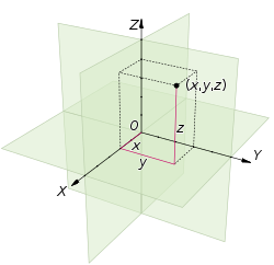
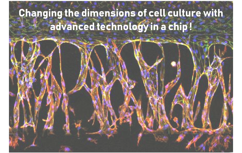
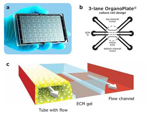
Authors Camilla Soragni and Gwenaëlle Rabussier are Early Stage Researchers located at MIMETAS, Netherlands. Read their previous blog post here. By definition, 3D modelling is the process of creating a three-dimensional model of an object. And what about dimensions? Curious to know how this conceptual representation emerged? Let’s go back approximately 570 BC, to its earliest development in Greek mathematics, with the Pythagoreans and their most famous theorem. The Pythagoreans’ use of one-dimensional lines laid down to describe a two-dimensional feature was one of the earliest examples of extending a single dimension in a way that creates an extra orthogonal dimension. In fact, a single line is considered to be one-dimensional, and a second line, non-parallel to the first is laid down and connect to any point of the first: a two-dimensional space is created.  Figure 1: Euclidian space Figure 1: Euclidian space Euclid, born around 325 BC, defines a point as “that which has no part” and defines a line as “breadthless point”, asserting the Pythagorean use of lines as one-dimensional objects. He also defines a surface, or 2-dimension, as that which has “length and breadth only”. He then indicated that a third spatial dimension exists specifically in relation to the first two spatial dimensions. He thus took the Pythagorean concept further and created a third dimension orthogonal to both the previous ones. [1] In the past decades, visualization, representation, and modeling in a 3-dimension space has been embraced by a wide variety of sectors. The main reason for its popularity lies in its power to provide excitement and more importantly, better understanding. Let us take the example of 3D printing. Who has never been excited about watching this technology in action? This undeniable enthusiasm is mainly due to its ability to bring 2D objects out of a computer screen into the physical world, enabling anyone to explore details in 3D reality, communicating complex information and inspiring genuine feeling to gain a fuller understanding of what is being shown. Its advantages are not limited to this. If this technology is widely used in manufacturing processes, it is also due to a large panel of benefits such as faster production, better quality, tangible design and product testing, risk reduction, to cite only a few [2]. And so, what about the use of 3D modeling in health science and cell biology? Interestingly, its benefits are pretty similar to those brought by 3D printing. Let us explain how. The indispensable tool to improve our perception and understanding of cell biology, development of tissue engineering, mechanism of diseases and drug action is cell culture. Cell culture is the process by which cells are grown under controlled conditions (e.g., essential nutrients, growth factors, gases), generally outside their environment. While 2D cell culture has traditionally been a very stable and simple method of growing cells, 3D cell culture is now bringing in vitro modeling to a higher level. Two-dimensional cell culture, which relies on the growth of cells on flat dishes, typically made of plastic, has been widely used since the 1900s and is still a dominant method in many biological studies. Even though 2D cell culture brought countless biological breakthroughs, these systems are not representative of real cell environments. Why? Simply because a flat surface is not a good way to understand how cells grow and function in a human body. It misses crucial features such as cell signaling, chemistry and complex organization. Yet, those are precisely characteristics of a 3-dimensional human body that we should model to get a better understanding of its biology. This is why over the past 10 years, the addition of a third dimension to 2D cell culture has received remarkable attention. Enabling complex interactions between cells as well as cells and the extracellular matrix (ECM), 3D cell culture retains the phenotypic and functional characteristics of their in vivo situations, providing a more physiologically relevant in vitro system to evaluate biological responses. If we take the example, Figure 2, of endothelium modeling with the culture of HUVEC (Human Umbilical Vascular Endothelial cells), you will only be able to grow a monolayer of endothelial cells in the conventional 2D culture, missing crucial cellular functions, while culture of these HUVECs within an ECM scaffold allows the formation of capillary-like microtissues which can closely mimic the functions of living tissues. [3] [4] [7] There are different ways of doing 3D cell culture. It goes from culture of cells into a 3D ECM scaffold (e.g., hydrogel) in which cells keep growing, maturing, ending up interacting with each other, to the formation of organoids, which are tiny, self-organized, multi-layered three-dimensional tissue cultures that are derived from stem cells (cells that can divide indefinitely and produce different types of cells as part of their progeny), replicating much of the complexity of an organ. But our personal favorite, which brought us to do our PhD in that field, is the Organ on-a-chip technology. Organs-on-chips are microfluidic devices in which cells or tissues are grown in a 3D manner, against or within an ECM scaffold. This combination of engineered three-dimensional tissue and microfluidic system enables us to stimulate the mechanics and physiology of organs. Since we are both PhD students at Mimetas – The Organ on-a-chip company, we will describe the principle of this technology with one of the platforms we are working with, the 3-lane OrganoPlate® (Fig. 3.a). What is thrilling about this microtechnology is the combination of connected multi-microchannels that enables compartmentalization and cell/ECM patterning (Fig 3.b). In addition to the advantages of 3D cell culture cited above, it allows the co-culture of different cell types and an increased control of their microenvironment. It provides a higher degree of structural complexity, such as tissue-tissue interface, and can thus better model how different types of cell interact. Another exciting feature is fluids flowing integration (Fig 3.c.). Like blood flooding into our blood-vessel, interstitial flow which supports cell differentiation and metabolism, is crucial for the functioning of all tissues and is one missing point of 2D cell culture. It also allows the improved and continuous transport of nutrients to cells leading to higher tissue viability. By replicating the human body more closely and better simulating mechanical, chemical and surface properties of the living organism, 3D cell culture, and especially 3D organ modeling in organ on-a-chip provides an exciting tool for a better understanding of organ complexity, mechanisms underlying diseases, drug screening and drug development. Their systematic use will help pharmaceutical industry save time and money by reducing drug trials periods while making them more precise or targeted and will consequently limit the number of animals destined to clinical testing. [5][6][7] Ever since 3D modelling emerged, the technology has not stopped evolving, and we are excited about what will be coming next… References
Leave a Reply. |
About the blogBeing a PhD student in a European training network is a life-changing adventure. Moving to a new country, carrying out a research project, facing scientific (and cultural) challenges, travelling around Europe and beyond… Those 3 years certainly do bring their part of new - sometimes frightening - but always enriching experiences. Categories
All
Archives
December 2021
|


 RSS Feed

5/2/2021
0 Comments