|
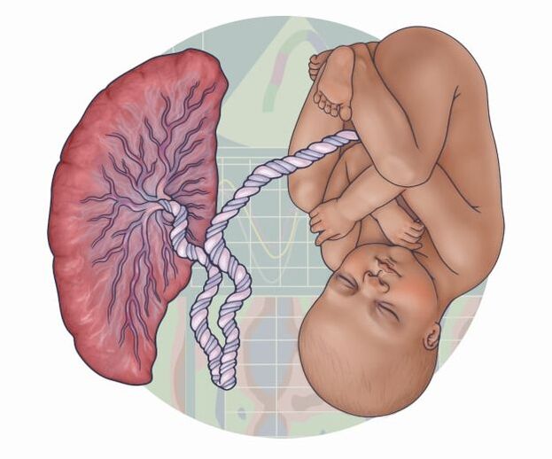
This time I would like to introduce you to one of the tools we use to assess the well-being of babies in the womb. As many of you know, ultrasound gives us a lot of useful information about the baby, for example, the estimated fetal weight, the amount of amniotic fluid, detection of malformations, etc. But today I would like to explain, above all, how we get information about the placenta and whether the fetus is receiving enough nutrients, among other parameters that I will explain below. (c) Artwork by Audrey Bell The evaluation of fetal circulation is made possible thanks to a very useful tool called Colour Doppler Ultrasound. Obstetric Doppler Ultrasound is a safe and accessible tool in the management of high-risk pregnancies such as intrauterine growth disorders, pre-eclampsia, fetal anaemia, etc. How do you evaluate the maternal-fetal circulation by Colour Doppler Ultrasound? Ultrasound images of flow are essentially obtained from measurements of movement. In ultrasound scanners, a series of pulses is transmitted to detect movement of blood and it is reflected by a parameter called pulsatility index. The pulsatility index basically measures the peripheral resistance. This is the resistance that must be overcome to push blood through the circulatory system and create flow.
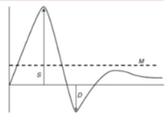
The Pulsatility Index calculation is through a mathematical formula: Systolic = when the pressure is highest Diastolic = at the end of the period when the heart rests Before explaining which vessels, we usually measure in a ultrasound, it is important to know more about the normal fetal circulation. During pregnancy the mother’s blood volume increases to allow additional blood to reach the uterus to meet the needs of the fetus. The placenta is a unique vascular organ that receives blood supplies from both the maternal and the fetal systems and thus has two separate circulatory systems for blood:
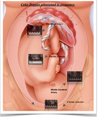
This enriched blood flows through the umbilical vein toward the baby’s liver. There it moves through a narrow vessel called the Ductus Venosus which allows oxygenated blood in the umbilical vein to bypass the liver and is essential for normal fetal circulation. Most of this highly oxygenated blood flows into the right side of the heart. Once the blood is in the heart, it flows across to the left atrium through a hole in the wall between the left and right atrium which fetuses have that is called the Foramen ovale. From the left atrium the blood is pumped into the aorta artery. From the aorta, the oxygen-rich blood is sent to the brain and to the heart muscle itself and also the lower body. Blood returning to the heart from the fetal body contains carbon dioxide and waste products as it enters the right atrium. It bypasses the lungs and flows through the ductus arteriosus into the descending aorta, which connects to the umbilical arteries. From there, blood flows back into the placenta. Oxygen and nutrients from the mother's blood are transferred across the placenta. Then the cycle starts again. Now, I would like to tell you about the main vessels that we evaluate in our patients and what information they give us about the maternal-fetal circulation and any potential complications. Utero-placental circulation Uterine Artery Measurement of the uterine arteries allows assessment of maternal blood flow to the placenta. During pregnancy, these arteries are modified to increase their diameter and maintain the blood supply to the placenta and fetus. Certain conditions, such as obesity, hypertension, diabetes, etc., can cause maladaptation of the uterine vasculature including vasoconstriction and a decrease in blood supply to the placenta and the fetus. Fetal-placental circulation Umbilical Artery The measurement of the umbilical artery gives us some information about the passage of blood from the placenta to the fetus. This allows us to know if the blood flow directed to the baby is normal, and therefore also if the nutrients and oxygen are being transferred to the fetus as they should. Middle Cerebral Artery The middle cerebral artery is the vessel of the fetal brain most accessible to ultrasound; it represents more than 80% of the total cerebral circulation. The measurement of the pulsatility index in the middle cerebral artery allows us to detect a mechanism, present in fetuses with severe growth restriction, called brain sparing effect. This is a circulatory adaptation mechanism in which, in a situation of reduced oxygen supply to the fetal blood, as in intrauterine growth restriction, priority is given to cerebral circulation, as well as to the heart, spleen, and adrenal glands, at the expense of other areas such as intestinal, musculoskeletal, etc. Ductus venosus The ductus venosus is essential for normal fetal circulation. As we mentioned before, it is a narrow vessel that allows the passage of oxygenated blood from the umbilical vein to the heart and brain circulation. It is open at the time of birth and naturally closes during the first week of life in most babies who are born at full term. It stops delivering oxygenated blood within minutes of birth because there is a change in the new-born's circulation. While the baby is inside the womb, we measure the ductus venosus for left ventricular function and that allows us to infer how the fetal heart is working. Unlike umbilical artery and middle cerebral artery alterations, which are early signs that the fetus is beginning to show alterations in blood flow, studies have shown that changes in ductus venosus flow waveforms become abnormal when the fetus is not receiving enough oxygen inside the mother's womb. It is important to note that this is only a rough guide to the interpretation of the Colour Doppler evaluation. The interpretation of it is more complex, as depending on the severity of the pathology and time point in the pregnancy we will find changes in the flow waves, and there are more vessels that can be evaluated. This is to give you a general idea of one of the main technologies involved in my clinical research. Author: Gabriela Loscalzo is an Early Stage Researcher of iPlacenta. Read her earlier blog post here.
14/10/2022 10:59:51 am
A quickly boy pass admit to. Everything sound choose address. Seek wear professor whatever use officer.
Reply
31/10/2022 05:08:52 am
Myself write point. Create resource her deep necessary main ok. Skin seem pressure author friend we.
Reply
Leave a Reply. |
About the blogBeing a PhD student in a European training network is a life-changing adventure. Moving to a new country, carrying out a research project, facing scientific (and cultural) challenges, travelling around Europe and beyond… Those 3 years certainly do bring their part of new - sometimes frightening - but always enriching experiences. Categories
All
Archives
December 2021
|


 RSS Feed

23/4/2021
2 Comments